

2023-11-03T02:48:52
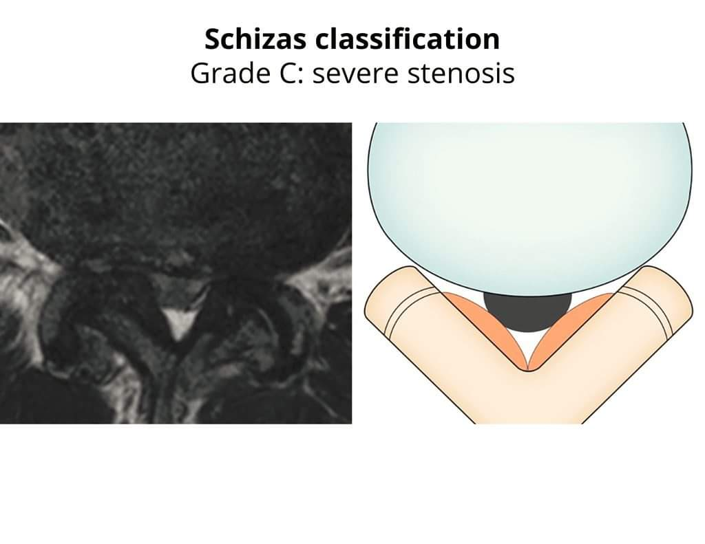
Physiotherapy clinic in Tambaram Are you Looking for Physiotherapy Treatment in Tambaram, Sunshine Super Speciality Physiotherapy Clinic, We Provide Electrotherapy, Exercise and Manual Therapy, Orthopedic, Neuro, Cardio, Pediatric, Sports and Geriatric Rehabilitation, Post Operative Physiotherapy Treatment, Fracture Rehabilitation, pain free movement. Radiographic Classification of Lumbar spinal stenosis💡 👉 Lumbar central canal stenosis (LCCS) is defined as the narrowing of the central spinal canal caused by degenerative changes with compression of neural and vascular structures, resulting in various degrees of clinical disability. https://pubmed.ncbi.nlm.nih.gov/21286714/ 👉 MRI plays a key role in the diagnosis of LCCS, and there have been reports of an inconsistent statistical association between MRI grading and the patient's disability or neurological impairment. https://pubmed.ncbi.nlm.nih.gov/27519125/, https://pubmed.ncbi.nlm.nih.gov/29939845/, https://pubmed.ncbi.nlm.nih.gov/31715409/, https://pubmed.ncbi.nlm.nih.gov/23830297/ 👉 Nevertheless, the assessment of LCCS using grading system is very widely used in clinical practices. https://pubmed.ncbi.nlm.nih.gov/32459814/, https://pubmed.ncbi.nlm.nih.gov/25079488/ 👉 One of the most prevalent radiographic classification systems of lumbar spinal stenosis war create by Schizas et al. https://pubmed.ncbi.nlm.nih.gov/20671589/. This system differentiates 7-grades based on the morphology of the dural sac on T2-weighted axial MRI with the rootlet/cerebrospinal fluid (CSF) fluid ratio taken into account: 1⃣ A1: the rootlets lie dorsally and occupy less than half of the dural sac area. 2⃣ A2: the rootlets lie dorsally, in contact with the dura but in a horseshoe configuration. 3⃣ A3: the rootlets lie dorsally and occupy more than half of the dural sac area. 4⃣ A4: the rootlets lie centrally and occupy the majority of the dural sac area. 5⃣ Grade B: the rootlets occupy the whole of the dural sac, but they can still be individualized. Some cerebrospinal fluid (CSF) is still present giving a grainy appearance to the sac. 6⃣ Grade C stenosis: no rootlets can be recognized, the dural sac demonstrating a homogeneous gray signal with no CSF signal visible. There is epidural fat present posteriorly. 7⃣ Grade D stenosis: no recognizable rootlets and no epidural fat posteriorly Illustrations: Case courtesy of Francis Deng, Radiopaedia.org. From the case rID: 92650 SUNSHINE ® SUPER SPECIALITY PHYSIOTHERAPY CLINIC - #DrParthiban #Sunshinephysioclinic.in #Physiotherapyclinicintambaram DR.M.P. PARTHIBAN.M.P.T (Ortho), Chief Orthopedic Physiotherapist, Call for Appointments: - 9345122177 East Tambaram, CHENNAI